Is Atrial Fibrillation Dangerous? 7 Serious Risks You Should Never Ignore
Is atrial fibrillation dangerous? Atrial fibrillation itself is rarely immediately life-threatening, but left untreated, it can lead to serious complications, including stroke, heart failure, and a higher risk of death. The good news is that with the right treatment, most of these risks can be greatly reduced.
Atrial fibrillation (AFib) is the most common heart rhythm disorder, affecting more than 10 million Americans, according to the American Heart Association. Some people feel a racing or fluttering heartbeat; others have no symptoms at all and find out only after a complication. Here are seven serious risks every person with atrial fibrillation should understand.
What Is Atrial Fibrillation?
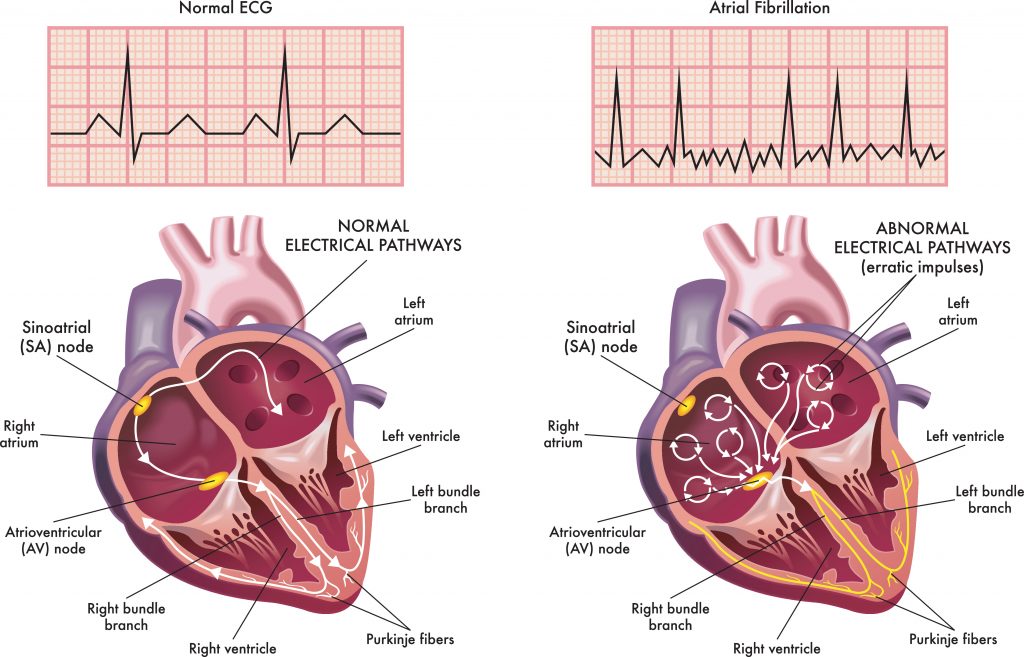
Normally, the heart’s upper chambers (atria) contract in a steady, coordinated rhythm. In atrial fibrillation, chaotic electrical signals cause the atria to quiver instead of beat effectively. The lower chambers (ventricles) often respond with a fast, irregular heartbeat.
Atrial fibrillation may come and go (paroxysmal), last longer (persistent) or become permanent. It tends to progress over time, which is why the 2023 ACC/AHA atrial fibrillation guideline treats it as a progressive disease and emphasizes early treatment.
1. Stroke and Blood Clots
Stroke is the most feared complication of atrial fibrillation. When the atria quiver instead of contracting, blood can pool and form clots, especially in a small vestigial pouch called the left atrial appendage. If a clot breaks loose, it can travel to the brain and cause a stroke, or block blood flow to the kidneys, intestines, or limbs (systemic embolism).
Atrial fibrillation raises the risk of stroke about fivefold. Strokes caused by atrial fibrillation also tend to be larger and more disabling than other strokes.
Your risk depends on other factors, including age, high blood pressure, diabetes, heart failure, prior stroke, vascular disease, and female sex. Cardiologists use a scoring system called CHA₂DS₂-VASc to estimate your risk and decide whether you need a blood thinner.
Know the signs of stroke (BE FAST): Balance loss, Eyes (vision changes), Face drooping, Arm weakness, Speech difficulty, Time to call 911.

2. Heart Failure
Atrial fibrillation and heart failure often feed each other. A fast, irregular heartbeat makes the heart pump less efficiently. When the atria quiver instead of contracting, the heart also loses the extra “kick” that helps fill the ventricles, which can reduce the amount of blood pumped by up to about 20% to 30% in some people.
Over time, this can cause or worsen heart failure, leading to shortness of breath, fatigue and swelling in the legs. People who have both conditions tend to have worse symptoms and outcomes than those with either one alone.
3. Tachycardia-Induced Cardiomyopathy
When atrial fibrillation keeps the heart racing for weeks or months, the heart muscle can become weakened and enlarged. This condition is called tachycardia-induced cardiomyopathy.
The encouraging news: it’s often reversible. When the heart rate is brought under control, or a normal rhythm is restored, heart function frequently improves, sometimes back to normal. That’s why it’s important to treat a persistently fast heart rate, even if you feel only mildly tired or short of breath.
4. Emergency Visits and Hospitalizations
Poorly controlled atrial fibrillation is a leading reason for emergency room visits and hospital admissions. Episodes of rapid heart rate, worsening heart failure, fainting, falls on blood thinners, and bleeding complications can all lead to hospital care.
Repeated hospitalizations disrupt life, increase health care costs and are often a sign that treatment needs to be adjusted. A clear treatment plan, including the right medications, possible rhythm control procedures, and close follow-up, can help keep you out of the hospital.
5. Cognitive Decline and Dementia
Research has linked atrial fibrillation to a higher risk of memory problems and dementia, even in people who have never had an obvious stroke. One reason is silent strokes: small areas of brain damage caused by tiny clots that don’t produce noticeable symptoms but add up over time. Reduced and irregular blood flow to the brain may also play a role.
An international report from the AF-SCREEN collaboration reviewed the growing evidence on atrial fibrillation and dementia. Appropriate blood thinners and good control of blood pressure and other risk factors are important steps to protect the brain.

6. Reduced Exercise Tolerance and Quality of Life
Even when atrial fibrillation isn’t dangerous in the moment, it can take a real toll on daily life. Common effects include:
- Fatigue and low energy
- Shortness of breath with activity
- Palpitations or a pounding heartbeat
- Dizziness or lightheadedness
- Anxiety about when the next episode will happen
- Trouble sleeping
Many people gradually cut back on activities they enjoy without realizing atrial fibrillation is the cause. Effective treatment often brings a dramatic improvement in energy and quality of life.
7. Higher Risk of Death
Atrial fibrillation is associated with a higher risk of death, and the AHA notes it can roughly double that risk. Much of this comes from the complications above, especially stroke and heart failure.
It’s important to keep this in perspective. Your individual outlook depends heavily on your overall heart health, other conditions such as high blood pressure, diabetes and sleep apnea, and whether your atrial fibrillation is properly treated. Many people with well-managed atrial fibrillation live long, active lives.
Symptoms of AFib and When to Call 911
Common symptoms include a fluttering, racing or irregular heartbeat, fatigue, shortness of breath, dizziness and chest discomfort. Some people, especially older adults, have no symptoms at all.
Call 911 if you have:
- Signs of stroke: face drooping, arm weakness, speech difficulty, sudden vision loss or loss of balance
- Chest pain or pressure
- Fainting or near-fainting
- Severe shortness of breath
- A very fast heartbeat with dizziness or weakness
For other emergency signs, see 7 life-threatening cardiac emergencies.
How Treatment Lowers Your AFib Risks
Modern atrial fibrillation care, as outlined in the 2023 guideline, rests on four main goals:
1. Preventing stroke. For people at elevated risk, blood thinners (anticoagulants) such as apixaban, rivaroxaban, dabigatran, edoxaban, or warfarin greatly reduce the risk of stroke. Aspirin is not an effective substitute. For people who can’t take blood thinners long term, left atrial appendage occlusion (such as the Watchman device) closes off the pouch where most clots form.
2. Controlling heart rate. Medications such as beta-blockers or calcium channel blockers slow a racing heart, relieve symptoms, and help prevent tachycardia-induced cardiomyopathy.
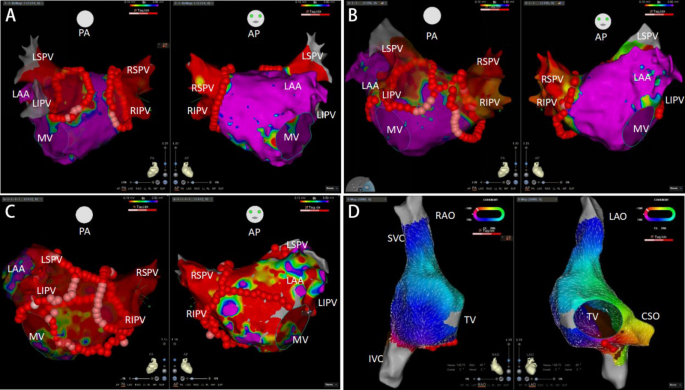
3. Restoring normal rhythm early. The guideline emphasizes early rhythm control. Options include antiarrhythmic medications, electrical cardioversion, and catheter ablation, which now has a Class 1 recommendation as first-line therapy for selected patients and for appropriate patients with heart failure with reduced ejection fraction.
4. Treating risk factors. Lifestyle and risk factor management is a core part of atrial fibrillation care:
- Weight loss if you have overweight or obesity
- Limit or avoid alcohol, a common atrial fibrillation trigger
- Treat sleep apnea
- Control blood pressure and diabetes. Learn how high blood pressure damages your body.
- Stay physically active. See heart-healthy exercises.
- Stop smoking and manage stress. Learn how stress affects your heart.

Frequently Asked Questions
Can you live a normal life with atrial fibrillation? Yes. With proper treatment, most people with atrial fibrillation live full, active lives. The keys are preventing stroke, controlling heart rate or rhythm, treating risk factors and keeping regular follow-up with your cardiologist.
Does atrial fibrillation go away on its own? Some episodes stop on their own, especially early on. But atrial fibrillation tends to return and progress over time. Even if your episodes stop, your stroke risk may remain, so don’t stop a blood thinner without talking to your doctor.
Do I need a blood thinner if I have atrial fibrillation? It depends on your stroke risk, not on how often you have atrial fibrillation. Your cardiologist will calculate your risk using a score such as CHA₂DS₂-VASc and weigh it against your bleeding risk. Many people with atrial fibrillation benefit from a blood thinner.
Can my smartwatch detect atrial fibrillation? Many smartwatches can detect an irregular rhythm and record an ECG. They’re helpful for spotting possible atrial fibrillation, and some are even FDA approved for the detection of atrial fibrillation, but a doctor should confirm the diagnosis. If your watch alerts you to atrial fibrillation, schedule an evaluation immediately.
Is atrial fibrillation caused by stress or caffeine? Stress, alcohol, poor sleep, and illness can trigger episodes in some people. Moderate caffeine is generally not a significant trigger for most people. The biggest drivers of atrial fibrillation are age, high blood pressure, obesity, sleep apnea, heart disease, and heavy alcohol use.
Atrial Fibrillation Care in Tampa
Atrial fibrillation is common, but its complications don’t have to be. Our board-certified cardiologist evaluates your stroke risk, heart function, and symptoms with in-office ECG, heart monitoring, and echocardiography, then builds a personalized plan to protect your heart and brain. When rhythm control procedures such as catheter ablation or left atrial appendage occlusion are recommended, we coordinate your care with electrophysiology colleagues at Tampa General Hospital and St. Joseph’s Hospital.
Not sure if your symptoms need a specialist? See the 10 warning signs you should see a cardiologist, and learn about advances in heart disease treatment.
Request an appointment or call (813) 540-4322.
602 S Audubon Ave, Suite A, Tampa, FL 33609
Medical Disclaimer
The content provided on the Ascent Cardiology website is intended for general educational and informational purposes only. The information, articles, and media presented on this website do not constitute medical advice, diagnosis, or treatment recommendations. Content published on this website is not intended to replace consultation, evaluation, or treatment by qualified healthcare professionals, including the providers at Ascent Cardiology or other licensed medical professionals.
If you believe you may be experiencing a medical emergency, call 911 or seek immediate emergency medical care.
The medical information, opinions, and educational materials presented on this website are provided for general awareness and may not reflect the specific clinical practices or medical opinions of Ascent Cardiology or its providers. Reliance on any information provided on this website is solely at your own risk.
This website may include links to third-party medical or educational websites for informational purposes only. Ascent Cardiology does not control, endorse, or assume responsibility for the accuracy, reliability, or content of any third-party websites linked from this site.
Sources
- What Is Atrial Fibrillation? — American Heart Association
- 2023 ACC/AHA/ACCP/HRS Guideline for the Diagnosis and Management of Atrial Fibrillation: Top Things to Know — American Heart Association
- 2023 Guideline for Diagnosis and Management of Atrial Fibrillation: Key Perspectives — American College of Cardiology
- Atrial Fibrillation and Dementia: A Report From the AF-SCREEN International Collaboration — Circulation

Heart Testing Before Organ Transplant: Things Every Patient Should Know
If you’re being evaluated for a kidney or liver transplant, you’ve probably been told you need “cardiac clearance.” For many

Autoimmune Diseases and Heart Disease: Hidden Risks You Should Know
If you live with lupus, rheumatoid arthritis, psoriasis, or another autoimmune condition, your care probably focuses on your joints, skin,

Varicose Vein Treatment: RFA vs. VenaSeal – 7 Things You Should Know
Aching, heavy legs. Swelling by the end of the day. Bulging, twisted veins you’d rather hide in shorts weather. If
Mitral Valve Disease: 7 Warning Signs You Should Never Ignore
Mitral valve disease often develops quietly. Many people live with a leaky or narrowed valve for years without knowing it, until breathlessness, fatigue, or an irregular heartbeat finally sends them to the doctor. By then, the heart may already be under strain.
The good news: mitral valve disease is easy to diagnose with a simple ultrasound, and treatment has advanced dramatically. Many patients can now have their valve repaired or replaced through a catheter, without open-heart surgery. Here’s how the mitral valve works, the seven warning signs to watch for, and your treatment options.
What Does the Mitral Valve Do?
Your heart has four valves that keep blood flowing in one direction. The mitral valve sits between the heart’s upper left chamber (left atrium) and lower left chamber (left ventricle). It opens to let oxygen-rich blood flow from the lungs into the left ventricle, then closes tightly so blood is pumped forward to the body.
When the mitral valve doesn’t open or close properly, the heart has to work harder, and pressure can back up into the lungs.

Types of Mitral Valve Disease
- Mitral regurgitation (leaky valve): The valve doesn’t close tightly, so blood leaks backward with each heartbeat. As the American Heart Association explains, blood moves in two directions with each beat. This is the most common form.
- Mitral valve prolapse: The valve’s flaps (leaflets) are floppy and bulge backward when the valve closes. Prolapse is common and usually harmless, but in some people it leads to significant leakage. Learn more about mitral valve prolapse.
- Mitral stenosis (narrowed valve): The valve becomes stiff and doesn’t open fully, restricting blood flow. Worldwide, it’s most often caused by rheumatic fever. In older adults in the U.S., it’s increasingly caused by calcium buildup around the valve.
Mitral regurgitation is also described by its cause. In primary (degenerative) regurgitation, the valve itself is damaged. In secondary (functional) regurgitation, the valve is normal but the heart muscle around it is enlarged or weakened, often from a heart attack or heart failure, which pulls the valve open.

7 Warning Signs of Mitral Valve Disease
Mild mitral valve disease often causes no symptoms. When symptoms appear, they usually develop gradually, so many people adjust their activity without realizing anything is wrong.
1. Shortness of Breath With Activity
Breathlessness is the most common symptom of mitral valve disease. You may notice it climbing stairs, carrying groceries, or walking at a pace that used to be easy. It happens because blood and pressure back up into the lungs.
2. Trouble Breathing When Lying Flat or at Night
Needing extra pillows to breathe comfortably, or waking up at night short of breath or coughing, is a sign that fluid may be collecting in the lungs. These symptoms suggest the valve problem is putting significant strain on the heart and should be evaluated promptly.
3. Unusual Fatigue and Lower Exercise Tolerance
When the heart can’t pump blood forward efficiently, your muscles and organs get less oxygen. Many patients describe feeling tired all the time, or notice they can’t do as much as they used to. Because it develops slowly, fatigue is often blamed on aging.
4. Heart Palpitations or an Irregular Heartbeat
A leaky or narrowed mitral valve can enlarge the left atrium, which can trigger atrial fibrillation (AFib), an irregular and often rapid heartbeat. You might feel fluttering, pounding or skipped beats. AFib raises the risk of blood clots and stroke, so new palpitations always deserve an evaluation.
5. Swelling in the Feet, Ankles, Legs or Abdomen
As mitral valve disease progresses, it can lead to heart failure and raise pressure in the lungs (pulmonary hypertension), which strains the right side of the heart. Fluid may build up in the lower legs or abdomen, and you may notice rapid weight gain.
6. A Heart Murmur
A murmur is an extra whooshing sound your doctor hears through a stethoscope. Mitral regurgitation and mitral stenosis each have a characteristic murmur. Many murmurs are harmless, but a new murmur, or one found along with symptoms, should be checked with an echocardiogram.
7. Lightheadedness, Chest Discomfort or Coughing
Some people with mitral valve disease feel dizzy or lightheaded, especially with exertion, or have chest discomfort. A persistent cough, sometimes with frothy or blood-tinged mucus, can occur when pressure in the lungs is high, particularly with mitral stenosis.
Call 911 for severe shortness of breath at rest, coughing up pink or frothy mucus, fainting, chest pain, or signs of stroke such as face drooping, arm weakness or trouble speaking. These can signal sudden worsening, such as a ruptured valve support cord, acute heart failure or a stroke from atrial fibrillation.
What Causes Mitral Valve Disease?
- Degenerative changes with age, including floppy or thickened valve leaflets and torn support cords (chordae)
- Mitral valve prolapse, which can run in families
- Heart attack or heart failure, which can stretch or distort the valve
- Calcium buildup around the valve in older adults, especially those with kidney disease
- Rheumatic heart disease, a complication of untreated strep throat
- Infective endocarditis, an infection of the heart valve
- Congenital heart conditions and certain connective tissue disorders
How Mitral Valve Disease Is Diagnosed
The key test is a transthoracic echocardiogram, a painless surface ultrasound of the heart. It shows how the valve opens and closes, how severe any leak or narrowing is, and how well the heart is pumping. Your cardiologist may also recommend:
- Transesophageal echocardiogram (TEE), which gives a much more detailed view of the valve from a probe passed into the esophagus, often used when planning repair or replacement
- ECG or heart monitor to check for atrial fibrillation
- Cardiac MRI to measure leakage and heart size precisely
- Exercise testing to see how symptoms and valve function change with activity
People with moderate or severe disease usually need echocardiograms every 6 to 12 months to watch for changes, because the best time to fix a valve is often before the heart muscle is permanently damaged.

Treatment Options for Mitral Valve Disease
Treatment depends on the type and severity of your valve problem, your symptoms, and your overall health. A heart team, including an interventional cardiologist and a cardiac surgeon, often reviews the options together.
Monitoring and medications. Mild disease may need only regular checkups and echocardiograms. Medications can relieve symptoms and treat related conditions, such as diuretics for fluid, heart failure medications for secondary regurgitation, and blood thinners for atrial fibrillation.
Surgical repair or replacement. For many people with primary mitral regurgitation, surgical repair is the preferred treatment, preserving the patient’s own valve. When repair isn’t possible, the valve can be replaced. Some centers offer minimally invasive or robotic surgery through small incisions.
Transcatheter edge-to-edge repair (TEER). For people who are at high risk for surgery, a clip (such as MitraClip or PASCAL) is delivered through a vein in the leg to hold the valve leaflets together and reduce leakage. In the COAPT trial of patients with heart failure and severe secondary regurgitation, TEER plus medication reduced death or heart failure hospitalization by 47% over five years compared with medication alone. Learn more about TEER from Cleveland Clinic.
Transcatheter mitral valve replacement (TMVR). This is the newest option. In December 2025, the FDA approved the SAPIEN M3, the first mitral valve replacement delivered entirely through a vein in the leg, for patients who aren’t candidates for surgery or TEER. Another device, Tendyne, approved earlier in 2025, is placed through a small incision in the chest.
Balloon valvuloplasty. For selected patients with rheumatic mitral stenosis, a balloon threaded through a catheter can widen the narrowed valve.
To learn more about catheter-based heart procedures, read our posts on the benefits of minimally invasive heart treatment and advances in heart disease treatment.
Frequently Asked Questions
Is mitral valve disease serious? It can be. Mild disease often stays stable for years and may only need monitoring. But moderate to severe disease can lead to heart failure, atrial fibrillation, stroke, and pulmonary hypertension if it isn’t treated at the right time. Regular echocardiograms help your cardiologist decide when to act.
Can mitral valve disease go away on its own? Structural valve damage doesn’t heal on its own. However, secondary mitral regurgitation sometimes improves when the underlying heart failure or atrial fibrillation is treated, because the heart becomes smaller and the valve closes better.
Is mitral valve prolapse dangerous? For most people, no. Mitral valve prolapse is common and usually causes little or no leakage. A small number of people develop significant regurgitation or heart rhythm problems over time, so periodic checkups with an echocardiogram are recommended.
Do I need open-heart surgery to fix my mitral valve? Not necessarily. Surgery remains the best option for many people, especially younger, healthier patients with primary regurgitation. For those at higher surgical risk, catheter-based options such as TEER or transcatheter valve replacement may be possible.
Can I exercise with mitral valve disease? Most people with mild disease can exercise normally. If your disease is moderate or severe, ask your cardiologist which activities are safe for you. See our guide to heart-healthy exercises.
Expert Heart Valve Evaluation in Tampa
At Ascent Cardiology, mitral valve disease is evaluated by a board-certified interventional cardiologist who is also a Fellow of the American Society of Echocardiography. We perform echocardiograms in our office, follow valve disease closely over time, and help you understand when treatment is needed. If a procedure is recommended, we coordinate care with the heart teams at Tampa General Hospital and St. Joseph’s Hospital.
If you’ve been told you have a murmur or a leaky valve, or you’re noticing new breathlessness or palpitations, don’t wait. See the 10 warning signs you should see a cardiologist.
Request an appointment or call (813) 540-4322.
602 S Audubon Ave, Suite A, Tampa, FL 33609
Medical Disclaimer
The content provided on the Ascent Cardiology website is intended for general educational and informational purposes only. The information, articles, and media presented on this website do not constitute medical advice, diagnosis, or treatment recommendations. Content published on this website is not intended to replace consultation, evaluation, or treatment by qualified healthcare professionals, including the providers at Ascent Cardiology or other licensed medical professionals.
If you believe you may be experiencing a medical emergency, call 911 or seek immediate emergency medical care.
The medical information, opinions, and educational materials presented on this website are provided for general awareness and may not reflect the specific clinical practices or medical opinions of Ascent Cardiology or its providers. Reliance on any information provided on this website is solely at your own risk.
This website may include links to third-party medical or educational websites for informational purposes only. Ascent Cardiology does not control, endorse, or assume responsibility for the accuracy, reliability, or content of any third-party websites linked from this site.
Sources
- Mitral Valve Regurgitation — American Heart Association
- Mitral Valve Prolapse — American Heart Association
- Mitral Valve Disease — Cleveland Clinic
- Heart Valve Diseases: Types — NHLBI
- TEER (Transcatheter Edge-to-Edge Repair) — Cleveland Clinic
- Minimally Invasive Mitral Valve Repair Reduces Hospitalizations and Deaths (COAPT 5-Year Results) — American College of Cardiology (2023)
- FDA Approves Sapien M3 for Transseptal Mitral Valve Replacement — TCTMD (December 2025)
Athlete’s Heart: Symptoms, Causes & When to Worry
Athlete’s heart is the normal way the heart adapts to years of intense training. A well-trained athlete’s heart is often larger, beats more slowly at rest and pumps more blood with each beat. These changes are a sign of fitness, not disease.
However, some of these normal changes can look like serious heart conditions on an ECG or echocardiogram. And rarely, a dangerous heart problem can hide behind what looks like athlete’s heart. Here’s what’s normal, what causes these changes, and when an athlete should see a cardiologist.
What Is Athlete’s Heart?
Athlete’s heart, also called athletic heart syndrome, describes structural and electrical changes in the heart caused by regular, intense exercise. It’s most common in people who train many hours a week, such as competitive endurance athletes, team-sport athletes and dedicated recreational athletes.
Athlete’s heart is not a disease and doesn’t need treatment. In most cases, the changes partly reverse within a few months after an athlete reduces training.
What Causes Athlete’s Heart?
During exercise, the heart pumps up to five or more times as much blood as it does at rest. Over time, it remodels to handle that workload more efficiently. The type of training shapes how the heart adapts:
- Endurance training (running, cycling, swimming, rowing) mainly increases the size of the heart’s chambers, so each beat moves more blood.
- Strength training (weightlifting, wrestling, throwing sports) mainly thickens the heart’s walls to handle short bursts of high pressure.
- Mixed sports such as basketball, hockey and soccer often produce a combination of both.
A larger, more efficient heart can move the same amount of blood with fewer beats. That’s why many athletes have a resting heart rate in the 40s or even lower.
Common Signs of Athlete’s Heart
Athlete’s heart usually causes no symptoms. It’s often noticed during a physical, a pre-participation screening or a test done for another reason. Normal findings include:
- A slow resting heart rate (sinus bradycardia), sometimes below 50 beats per minute
- A heart murmur caused by increased blood flow, often harmless
- An enlarged heart on an echocardiogram or chest X-ray, with normal pumping function
- Mildly thickened heart walls
- ECG changes that would be unusual in non-athletes but are considered normal in trained athletes
Expert guidelines, known as the International Criteria for ECG interpretation in athletes, list ECG findings that are normal in athletes. These include a slow heart rate, mild heart block (first-degree AV block), early repolarization and high voltage suggesting a larger heart. Recognizing these patterns prevents athletes from being wrongly disqualified from sport.

Athlete’s Heart vs. Hypertrophic Cardiomyopathy and Other Conditions
The biggest challenge is telling athlete’s heart apart from heart conditions that can cause sudden cardiac arrest during exercise. The most important is hypertrophic cardiomyopathy (HCM), an inherited thickening of the heart muscle. We cover HCM in more detail in 5 lesser-known heart conditions.
Other conditions that can mimic or hide behind athlete’s heart include:
- Arrhythmogenic cardiomyopathy (ARVC), in which heart muscle is replaced by scar and fat
- Dilated cardiomyopathy, a weakened, enlarged heart
- Myocarditis, inflammation of the heart muscle, often after a viral illness
- Coronary artery anomalies, arteries that start or run in an abnormal location
- Inherited rhythm disorders, such as long QT syndrome, Brugada syndrome and Wolff-Parkinson-White syndrome
In athletes over 35, coronary artery disease becomes the most common cause of exercise-related cardiac events. The ACC summarizes the causes of sudden cardiac arrest and death in athletes.
When to Worry: Warning Signs in Athletes
Athlete’s heart itself doesn’t cause symptoms. If an athlete has any of the following, they should stop training and see a cardiologist:
- Fainting or near-fainting during or right after exercise. This is the most important red flag.
- Chest pain or pressure with exertion
- Shortness of breath out of proportion to the effort
- Palpitations, a racing or irregular heartbeat, especially with dizziness
- A sudden drop in performance or unusual fatigue without explanation
- Symptoms after a viral illness, such as chest pain or palpitations after COVID-19 or the flu
- A family history of sudden cardiac death before age 50, cardiomyopathy or inherited rhythm disorders
- An abnormal ECG flagged during a screening physical
Long-term endurance athletes, especially middle-aged men, also have a higher risk of atrial fibrillation, an irregular heart rhythm. The ACC reviews what contributes to AFib in former endurance athletes. For a broader checklist of symptoms, see 10 warning signs you should see a cardiologist.
Call 911 if an athlete collapses. Start CPR and use an AED immediately. Every minute without CPR and defibrillation lowers the chance of survival.
How Doctors Diagnose Athlete’s Heart
A cardiologist experienced with athletes uses several tools to separate normal adaptation from disease:
- 12-lead ECG, interpreted using athlete-specific criteria
- Echocardiogram to measure wall thickness, chamber size, pumping and filling
- Cardiac MRI to look for scarring or abnormal thickening patterns, especially in gray-zone cases
- Cardiopulmonary exercise testing to measure fitness and check rhythm during exercise
- Holter or event monitoring to record the heart’s rhythm over days to weeks
- Genetic testing when an inherited condition is suspected
- Supervised detraining, a short period of reduced exercise, followed by repeat imaging
The goal is to keep healthy athletes in their sport while protecting those with a real risk. Today, many athletes with a heart condition can continue to compete after an individualized, shared decision-making discussion with their cardiologist.
Heart Screening for Athletes
In the U.S., pre-participation physicals typically include a medical history, family history, and physical exam. Some programs, including many collegiate and professional leagues, also add an ECG and echocardiogram. Beginning in the 2026-2027 school year, under Florida’s Second Chance Act (Florida Statute 1006.20), students participating in any Florida High School Athletic Association (FHSAA)-sanctioned athletic tryout, conditioning, practice, or competition for the first time must have a cleared electrocardiogram (ECG/EKG) before they can participate.
Read the American Heart Association’s overview of screening young athletes for heart disease.
Adult and masters athletes, especially those over 35 or with risk factors such as high blood pressure, high cholesterol, diabetes or smoking, may benefit from a cardiology evaluation before starting or intensifying training.
Frequently Asked Questions
Is athlete’s heart dangerous? No. Athlete’s heart is a normal, healthy adaptation to training. The concern is making sure it isn’t hiding a different heart condition, which a cardiologist experienced with athletes can sort out.
Is a resting heart rate in the 40s normal for athletes? Yes. Trained athletes often have resting heart rates in the 40s and sometimes lower. A slow heart rate is a concern only if it comes with dizziness, fainting, fatigue or shortness of breath.
Does athlete’s heart go away? The changes usually partly reverse within a few months of reducing training. This is one way doctors distinguish athlete’s heart from conditions like hypertrophic cardiomyopathy, which don’t improve with rest.
Can you have athlete’s heart without being a professional athlete? Yes. Marathon runners, triathletes, cyclists and other dedicated recreational athletes who train many hours a week can develop athlete’s heart.
Should my child get an ECG before playing sports? It depends on your child’s history, symptoms, family history and sport. A doctor can review whether an ECG is appropriate. Any child who faints during exercise, has chest pain with exertion or has a family history of sudden cardiac death should see a cardiologist before playing.
Sports Cardiology Care in Tampa
As team cardiologist for the Tampa Bay Lightning and former team cardiologist for the Toronto Raptors, our cardiologist has evaluated elite athletes at the highest level of competition. We bring the same expertise to athletes of every age and level, from high school competitors to weekend warriors and masters athletes.
We offer athlete-specific ECG interpretation, echocardiography, exercise testing and individualized return-to-play guidance.
Request an appointment or call (813) 540-4322.
602 S Audubon Ave, Suite A, Tampa, FL 33609
Medical Disclaimer
The content provided on the Ascent Cardiology website is intended for general educational and informational purposes only. The information, articles, and media presented on this website do not constitute medical advice, diagnosis, or treatment recommendations. Content published on this website is not intended to replace consultation, evaluation, or treatment by qualified healthcare professionals, including the providers at Ascent Cardiology or other licensed medical professionals.
If you believe you may be experiencing a medical emergency, call 911 or seek immediate emergency medical care.
The medical information, opinions, and educational materials presented on this website are provided for general awareness and may not reflect the specific clinical practices or medical opinions of Ascent Cardiology or its providers. Reliance on any information provided on this website is solely at your own risk.
This website may include links to third-party medical or educational websites for informational purposes only. Ascent Cardiology does not control, endorse, or assume responsibility for the accuracy, reliability, or content of any third-party websites linked from this site.
Sources
- Athlete’s Heart — Cleveland Clinic
- International Recommendations for Electrocardiographic Interpretation in Athletes — Journal of the American College of Cardiology
- Sudden Cardiac Arrest and Death in Athletes: Key Points — American College of Cardiology
- Differential Diagnosis Between Athlete’s Heart and Cardiomyopathies — NIH/PMC
- What Contributes to a High Prevalence of AFib in Former Elite Endurance Athletes? — American College of Cardiology
- Screening Young Athletes for Heart Disease — American Heart Association

Heart Testing Before Organ Transplant: Things Every Patient Should Know
If you’re being evaluated for a kidney or liver transplant, you’ve probably been told you need “cardiac clearance.” For many

Autoimmune Diseases and Heart Disease: Hidden Risks You Should Know
If you live with lupus, rheumatoid arthritis, psoriasis, or another autoimmune condition, your care probably focuses on your joints, skin,

Varicose Vein Treatment: RFA vs. VenaSeal – 7 Things You Should Know
Aching, heavy legs. Swelling by the end of the day. Bulging, twisted veins you’d rather hide in shorts weather. If
7 Advances in Heart Disease Treatment You Should Know About
Recent advances in heart disease treatment have transformed care in the past decade. Procedures that once required open-heart surgery can now be done through small catheters and minimally invasive techniques. Medications have extended and improved the lives of people with heart failure. In addition, new imaging can find heart disease earlier and more accurately than ever.
Here are seven advances in heart disease treatment that are changing outcomes for patients today, and what each could mean for you.
1. Modern Drug-Eluting Coronary Stents
A stent is a tiny mesh tube placed in a narrowed heart artery to keep it open. Early bare-metal stents often caused the artery to narrow again. Today’s drug-eluting stents slowly release medication that prevents scar tissue from growing inside the stent.
The newest generation uses thinner struts and coatings that dissolve over time. As a result, rates of re-narrowing and stent clotting are now low. Many patients can also take two blood thinners for a shorter time than in the past, which lowers bleeding risk. Researchers have found that, for some patients, one month of dual antiplatelet therapy can be safe after stent placement.
2. Intravascular Lithotripsy for Calcified Coronary Arteries
As people age, plaque in the heart arteries can harden with calcium, almost like bone. Coronary artery calcification makes it difficult to open the artery fully and expand a stent properly.
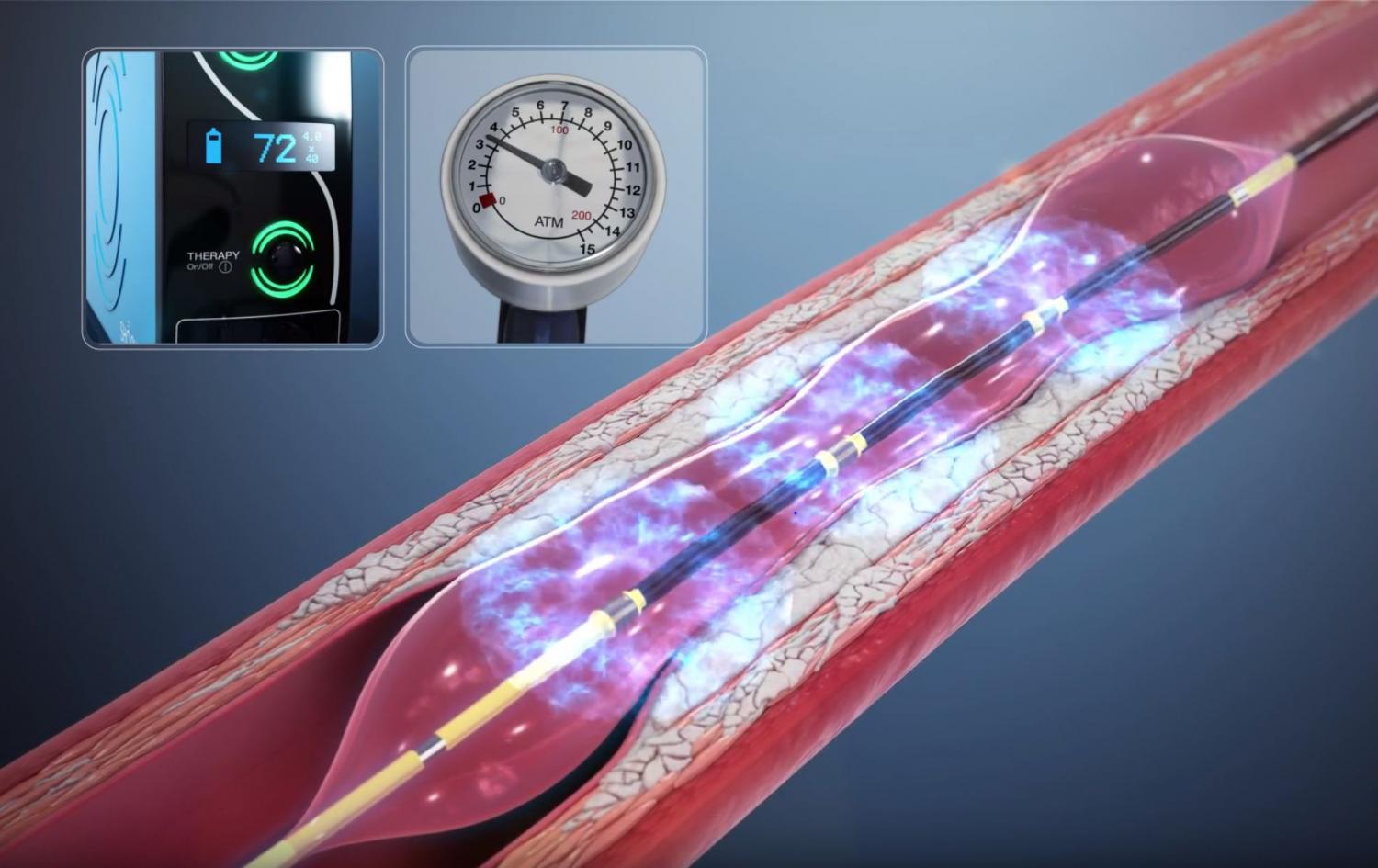
Intravascular lithotripsy (IVL) borrows technology used to break up kidney stones. A special balloon delivers sound-based pressure waves that crack the calcium inside the artery wall. The artery can then be opened and the stent fully expanded, with low rates of complications. For many patients with heavily calcified arteries, IVL has made catheter-based treatment possible where surgery once seemed like the only option. The American College of Cardiology reviews how IVL is used in contemporary practice.
3. Precision-Guided PCI With IVUS and OCT
Traditional angiograms show the outline of the artery on an X-ray, but not the artery wall itself. Intravascular imaging changes that:
- Intravascular ultrasound (IVUS) uses sound waves from a tiny probe inside the artery to measure plaque, vessel size and calcium.
- Optical coherence tomography (OCT) uses light to create very high-resolution images of the artery wall and the stent.
With these tools, cardiologists choose the right stent size, confirm it is fully expanded and check that its edges are well positioned. Studies show imaging-guided PCI reduces repeat procedures and heart events compared with angiography alone, particularly for complex blockages. Read the American College of Cardiology summary. For more on how these procedures help patients, see our post on the benefits of minimally invasive heart treatment.
4. Transcatheter Heart Valve Replacement (TAVR)
Aortic stenosis is a narrowing of the aortic valve that makes the heart work harder and can cause breathlessness, chest pain and fainting. In the past, the only replacement option was open-heart surgery.
With transcatheter aortic valve replacement (TAVR), a new valve is delivered through a catheter, usually from the groin, and placed inside the diseased valve. TAVR is now approved for patients across the full range of surgical risk. Many go home within one to two days. A heart team that includes an interventional cardiologist and a cardiac surgeon decides whether TAVR or surgery is the better option for each patient. Learn more about TAVR from NHLBI.
5. Newer Heart Failure Medications
Heart failure means the heart isn’t pumping or filling as well as it should. For people with a weakened heart muscle (heart failure with reduced ejection fraction), guidelines now recommend four classes of medication, often called the “four pillars”:
- ARNI therapy (sacubitril/valsartan, which relaxes blood vessel and reduces strain on the heart
- Beta-blockers, which slow the heart rate and protect the heart muscle
- Mineralicorticoid receptor antagonists (spironolactone, finerenone), which reduce fluid buildup and scarring
- SGLT2-inhibitors (such as dapagliflozin and empagliflozin, originally diabetes medications, that also protect the heart and kidneys, even in people without diabetes
Together, these medications lower the risk of hospitalization and death and can help the heart muscle recover. SGLT2-inhibitors also benefit many patients with heart failure with preserved ejection fraction, a form that once had few proven treatments. See the AHA’s guide to medications used to treat heart failure and the ACC’s key points from the heart failure guideline.

6. Coronary CT Angiography and CT-Derived FFR
Coronary CT angiography (CCTA) uses a fast CT scan and contrast dye to create detailed 3D images of the heart arteries, without a catheter. It can show both hard (calcified) and soft plaque, so heart disease can be found before it causes a heart attack.
CT-derived fractional flow reserve (FFR-CT) takes this further. Computer modeling of the CT images estimates whether a narrowing actually reduces blood flow. This helps identify who truly needs a catheter procedure and who can be treated with medication alone. National chest pain guidelines now support CCTA as a first-line test for many people with chest pain. The ACC reports that CT scanning is highly effective for evaluating chest pain. Some chest pain comes from conditions a CT scan can miss, such as microvascular disease, which we cover in 5 lesser-known heart conditions.

7. Leadless Pacemakers and Advanced Cardiac Devices
Traditional pacemakers sit under the skin of the chest and connect to the heart with wires called leads. Over time, leads and the pocket under the skin can cause problems such as infection or lead fracture.
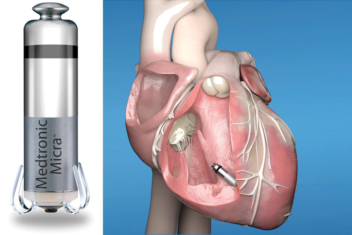
A leadless pacemaker is a self-contained device about the size of a large vitamin. It’s placed directly inside the heart through a catheter in the leg, so there’s no chest incision, no visible bump and no leads. Newer systems, such as the FDA-approved Aveir leadless pacing system, can now pace more than one chamber of the heart.
Other device advances include defibrillators that don’t need wires inside the heart and implantable monitors that track heart rhythm for years.
What These Advances in Heart Disease Treatment Mean for You
These advances give patients more options, faster recovery and more personalized care. However, the newest treatment isn’t always the right one. The best plan depends on your symptoms, test results, other health conditions and personal goals.
If you have symptoms such as chest pain, shortness of breath, palpitations or fainting, don’t wait. Review the 10 warning signs you should see a cardiologist and schedule an evaluation.
Frequently Asked Questions
Are drug-eluting stents safe long-term? Yes. Modern drug-eluting stents have strong long-term safety records and low rates of re-narrowing. Taking your prescribed blood thinners and controlling cholesterol, blood pressure and diabetes helps keep them working.
Who is a candidate for TAVR? TAVR is approved for people with severe aortic stenosis across all levels of surgical risk. A heart team considers your age, anatomy, overall health and preferences to decide between TAVR and surgery.
Can heart failure improve with medication? Yes. With today’s guideline-recommended medications, many people with heart failure feel better, have fewer hospital stays and see their heart function improve, sometimes significantly.
Is a coronary CT angiogram better than a stress test? They answer different questions. A coronary CT angiogram shows the arteries and plaque directly, while a stress test shows how the heart performs under exertion. Your cardiologist will recommend the right test for your situation.
How long does a leadless pacemaker battery last? Battery life depends on how often the device paces, but it typically lasts many years. When the battery runs low, a new device can usually be added or the old one retrieved, depending on the system.
Access Advanced Heart Care in Tampa
Our board-certified interventional cardiologist offers modern, evidence-based heart care, from advanced imaging and echocardiography to catheter-based procedures at Tampa General Hospital and St. Joseph’s Hospital. We’ll explain your options clearly and build a plan around you.
Request an appointment or call (813) 540-4322.
602 S Audubon Ave, Suite A, Tampa, FL 33609
Medical Disclaimer
The content provided on the Ascent Cardiology website is intended for general educational and informational purposes only. The information, articles, and media presented on this website do not constitute medical advice, diagnosis, or treatment recommendations. Content published on this website is not intended to replace consultation, evaluation, or treatment by qualified healthcare professionals, including the providers at Ascent Cardiology or other licensed medical professionals.
If you believe you may be experiencing a medical emergency, call 911 or seek immediate emergency medical care.
The medical information, opinions, and educational materials presented on this website are provided for general awareness and may not reflect the specific clinical practices or medical opinions of Ascent Cardiology or its providers. Reliance on any information provided on this website is solely at your own risk.
This website may include links to third-party medical or educational websites for informational purposes only. Ascent Cardiology does not control, endorse, or assume responsibility for the accuracy, reliability, or content of any third-party websites linked from this site.
Sources
- What Is a Stent? — American Heart Association
- One Month of Dual Antiplatelet Therapy Is Safe and Feasible After Stent Placement — AHA Newsroom
- Coronary Artery Calcification — Cleveland Clinic
- Shocking the Rock With Coronary Intravascular Lithotripsy in Contemporary Practice — American College of Cardiology
- Intravascular Imaging-Guided PCI vs. Angiography-Guided PCI — American College of Cardiology
- What Is TAVR? — American Heart Association
- Transcatheter Aortic Valve Replacement — NHLBI
- Medications Used to Treat Heart Failure — American Heart Association
- 2022 AHA/ACC/HFSA Heart Failure Guideline: Key Perspectives — American College of Cardiology
- CT Scan Is Most Effective to Assess People With Chest Pain — American College of Cardiology
- Is a Leadless Pacemaker Right for You? — Cleveland Clinic
- Aveir Leadless Pacing System — U.S. Food and Drug Administration

Heart Testing Before Organ Transplant: Things Every Patient Should Know
If you’re being evaluated for a kidney or liver transplant, you’ve probably been told you need “cardiac clearance.” For many

Autoimmune Diseases and Heart Disease: Hidden Risks You Should Know
If you live with lupus, rheumatoid arthritis, psoriasis, or another autoimmune condition, your care probably focuses on your joints, skin,

Varicose Vein Treatment: RFA vs. VenaSeal – 7 Things You Should Know
Aching, heavy legs. Swelling by the end of the day. Bulging, twisted veins you’d rather hide in shorts weather. If
7 Benefits of Minimally Invasive Heart Treatment
Not long ago, treating a blocked heart artery or a failing valve almost always meant open-heart surgery. Today, minimally invasive heart treatment lets interventional cardiologists treat many of these conditions through a tiny puncture in the wrist or groin. Patients often go home the same day or the next morning.
The most common of these procedures is percutaneous coronary intervention (PCI), also called angioplasty and stenting. Other catheter-based treatments include transcatheter valve replacement and procedures for complex blockages. Here are seven benefits of this approach and what they mean for you.
1. Minimally Invasive Treatment Without Open-Heart Surgery
Traditional bypass surgery requires opening the breastbone and often stopping the heart. In contrast, minimally invasive heart treatment is performed through a thin, flexible tube called a catheter. The catheter is guided through an artery in the wrist (radial artery) or upper thigh (femoral artery) to the heart.
Most patients receive local anesthesia and light sedation rather than general anesthesia. There’s no large incision, no cut breastbone and no heart-lung machine. As a result, the procedure is easier on the body, especially for older adults and people with other health conditions. Learn what happens during a cardiac catheterization from the Society for Cardiovascular Angiography and Interventions.
2. Faster Recovery and Shorter Hospital Stays
Recovery is one of the biggest differences between catheter-based treatment and surgery.
- After angioplasty and stenting: Many patients go home the same day or after one night. Most return to normal activities within about a week.
- After open-heart surgery: A hospital stay of several days is typical, and full recovery often takes 6 to 12 weeks.
Wrist (radial) access has made recovery even easier. Compared with groin access, it lowers bleeding risk and lets patients sit up and walk soon after the procedure. See Cleveland Clinic’s guide to recovering after angioplasty and stenting.
3. Improved Blood Flow to the Heart
Coronary artery disease narrows the arteries that supply the heart with oxygen-rich blood. During angioplasty, a small balloon opens the narrowed artery. Then a stent, a tiny mesh tube, is placed to hold it open. Modern drug-eluting stents slowly release medicine that helps keep the artery from narrowing again.
Restoring blood flow lets the heart muscle get the oxygen it needs, especially during exercise. For patients with severe or high-risk blockages, this can also protect heart function over time.
4. Life-Saving Treatment During a Heart Attack
During a major heart attack, a blood clot completely blocks a coronary artery and heart muscle begins to die within minutes. Emergency angioplasty, known as primary PCI, is the preferred way to reopen the artery quickly. National guidelines aim to open the artery within 90 minutes of arriving at the hospital.
The faster blood flow returns, the more heart muscle is saved. That leads to better long-term heart function and survival. Read more about heart attack treatment from the American Heart Association. Women’s heart attack symptoms can be subtle, so also review our guide to women’s heart attack symptoms.
5. Relief of Angina and Improved Quality of Life
Angina is chest pain or pressure caused by reduced blood flow to the heart. It can make walking, climbing stairs or exercising uncomfortable or frightening. Medications are the first step for stable angina. However, when symptoms continue despite medication, angioplasty and stenting can provide significant relief.
Many patients find they can walk farther, return to activities they had given up, and rely on fewer medications for chest pain. Learn more about angina treatment options from NHLBI.
6. Advanced Precision With Modern Imaging Technology
Today’s interventional cardiologists don’t rely on X-ray pictures alone. Advanced tools show the inside of the artery and measure how much a blockage actually limits blood flow:
- Intravascular ultrasound (IVUS) and optical coherence tomography (OCT) show the artery wall from the inside, so stents can be sized and positioned precisely.
- Fractional flow reserve (FFR) and related pressure measurements show whether a narrowing is truly reducing blood flow, which helps avoid unnecessary stents.
For complex blockages, imaging-guided PCI has been linked to fewer repeat procedures and better outcomes than angiography alone. Read a summary of intravascular imaging-guided PCI research from the American College of Cardiology.
7. Individualized Treatment for Complex Cardiovascular Conditions
Minimally invasive heart treatment is no longer only for simple blockages. Catheter-based options now include:
- Complex PCI for heavily calcified arteries, blockages where arteries branch, and chronic total occlusions
- Transcatheter aortic valve replacement (TAVR) for severe aortic stenosis
- Peripheral interventions to restore blood flow to the legs
No single treatment is right for everyone. Your cardiologist will weigh your symptoms, anatomy, other health conditions and goals. For some patients, medication alone or bypass surgery is the better choice. When surgery may be the right option, an interventional cardiologist works with a cardiac surgeon as part of a heart team.
Who Is a Candidate for Minimally Invasive Heart Treatment?
You may be a candidate if you have:
- Chest pain or shortness of breath that continues despite medication
- An abnormal stress test or coronary CT scan showing significant blockages
- A heart attack or unstable angina
- Severe aortic stenosis, which may be treated with TAVR
- Leg pain when walking caused by blocked arteries
The first step is a thorough evaluation, which may include a stress test, echocardiogram, coronary CT angiography or diagnostic catheterization. Not sure whether your symptoms need attention? Read 10 warning signs you should see a cardiologist. Some chest pain comes from conditions that don’t need a stent, such as coronary microvascular dysfunction. Learn about these in our post on lesser-known heart conditions.
Frequently Asked Questions
Is angioplasty painful? Most patients feel only a brief pinch when the numbing medicine is given at the wrist or groin. You’ll be awake but relaxed with light sedation. Some people feel brief chest pressure while the balloon is inflated.
How long does a stent last? Stents are designed to stay in place permanently. Modern drug-eluting stents have low rates of re-narrowing. Taking your prescribed blood thinners and managing cholesterol, blood pressure and diabetes helps protect the stent and your other arteries.
How soon can I return to work after a stent? Many people with desk jobs return within a few days to a week after a planned procedure. Recovery after a heart attack usually takes longer, and cardiac rehabilitation is recommended.
Is minimally invasive heart treatment safe? Angioplasty and stenting are among the most commonly performed heart procedures, and serious complications are uncommon. Your cardiologist will review your individual risks and benefits before the procedure.
When is open-heart surgery the better choice? Bypass surgery may be better for some patients with blockages in several arteries, particularly those with diabetes or reduced heart function. Your cardiologist and a cardiac surgeon can review your case together to recommend the best option.
Explore Minimally Invasive Heart Treatment in Tampa
Our board-certified interventional cardiologist performs advanced, catheter-based heart procedures at Tampa General Hospital and St. Joseph’s Hospital. Every treatment plan starts with a thorough evaluation and a clear explanation of your options.
Request an appointment or call (813) 540-4322.
602 S Audubon Ave, Suite A, Tampa, FL 33609
Medical Disclaimer
The content provided on the Ascent Cardiology website is intended for general educational and informational purposes only. The information, articles, and media presented on this website do not constitute medical advice, diagnosis, or treatment recommendations. Content published on this website is not intended to replace consultation, evaluation, or treatment by qualified healthcare professionals, including the providers at Ascent Cardiology or other licensed medical professionals.
If you believe you may be experiencing a medical emergency, call 911 or seek immediate emergency medical care.
The medical information, opinions, and educational materials presented on this website are provided for general awareness and may not reflect the specific clinical practices or medical opinions of Ascent Cardiology or its providers. Reliance on any information provided on this website is solely at your own risk.
This website may include links to third-party medical or educational websites for informational purposes only. Ascent Cardiology does not control, endorse, or assume responsibility for the accuracy, reliability, or content of any third-party websites linked from this site.
Sources
- Percutaneous Coronary Intervention — NHLBI
- Stents: Getting a Stent — NHLBI
- Angina Treatment — NHLBI
- Angiogram/Cardiac Catheterization — SCAI SecondsCount
- After Your Interventional Procedure — Cleveland Clinic
- Heart Attack Treatment — American Heart Association
- Angina (Chest Pain) — American Heart Association
- What Is TAVR? — American Heart Association
- Intravascular Imaging-Guided PCI vs. Angiography-Guided PCI — American College of Cardiology

Heart Testing Before Organ Transplant: Things Every Patient Should Know
If you’re being evaluated for a kidney or liver transplant, you’ve probably been told you need “cardiac clearance.” For many

Autoimmune Diseases and Heart Disease: Hidden Risks You Should Know
If you live with lupus, rheumatoid arthritis, psoriasis, or another autoimmune condition, your care probably focuses on your joints, skin,

Varicose Vein Treatment: RFA vs. VenaSeal – 7 Things You Should Know
Aching, heavy legs. Swelling by the end of the day. Bulging, twisted veins you’d rather hide in shorts weather. If
5 Lesser-Known Heart Conditions: Symptoms You Shouldn’t Ignore
When most people think of heart disease, they picture blocked arteries and heart attacks. However, several lesser-known heart conditions cause real symptoms and are often missed for years. Some don’t show up on routine tests. Others mimic common problems like anxiety, asthma or aging.
Knowing these conditions can help you speak up when something doesn’t feel right. Here are five worth knowing, the symptoms to watch for, and how they’re diagnosed and treated.
1. Coronary Microvascular Dysfunction (CMD)
What it is: CMD affects the heart’s tiniest blood vessels rather than the large coronary arteries. These small vessels don’t widen properly, so the heart muscle doesn’t get enough blood during exertion or stress. Because the large arteries look normal, patients are often told their heart is “fine.”
Symptoms to watch for:
- Chest pain or pressure, often lasting longer than typical angina
- Chest pain during daily activities or emotional stress
- Shortness of breath and fatigue
- Trouble sleeping
Who’s at risk: CMD is more common in women, especially around and after menopause. High blood pressure, diabetes and high cholesterol also raise risk. Women’s heart symptoms often differ from men’s, so read our guide to women’s heart attack symptoms.
Diagnosis and treatment: CMD is diagnosed with specialized tests, including cardiac PET, stress cardiac MRI or coronary function testing in the cath lab. Treatment typically includes medications to relax blood vessels and control blood pressure, plus statins and lifestyle changes. Learn more about coronary microvascular disease from the American Heart Association.
2. Takotsubo Cardiomyopathy (Broken Heart Syndrome)
What it is: Takotsubo cardiomyopathy is a sudden, temporary weakening of the heart muscle. It is usually triggered by intense emotional or physical stress, such as the death of a loved one, a frightening event or a serious illness. A surge of stress hormones “stuns” the heart, and the main pumping chamber balloons out at the tip.
Symptoms to watch for:
- Sudden chest pain
- Shortness of breath
- Symptoms that look exactly like a heart attack
Who’s at risk: About 90% of cases occur in women, most often after menopause.
Diagnosis and treatment: Because broken heart syndrome mimics a heart attack, it needs emergency care. Doctors confirm it with a cardiac catheterization showing no major blockages, along with an echocardiogram showing the typical shape. Most people recover heart function within weeks with medications and monitoring. Read more about broken heart syndrome from Harvard Health.
3. Hypertrophic Cardiomyopathy (HCM)
What it is: HCM is an inherited condition in which the heart muscle becomes abnormally thick. The thickened muscle can block blood flow out of the heart and make it stiff. HCM affects about 1 in 500 people, yet many don’t know they have it.
Symptoms to watch for:
- Shortness of breath, especially with exercise
- Chest pain
- Dizziness or fainting, particularly during or right after exercise
- Palpitations
- A heart murmur found on exam
Who’s at risk: Anyone with a parent, sibling or child with HCM, since each child of an affected parent has a 50% chance of inheriting it. HCM is also a leading cause of sudden cardiac death in young athletes.
Diagnosis and treatment: An echocardiogram is the key test, often with cardiac MRI and genetic testing. Treatment has advanced significantly. Options include beta blockers, newer cardiac myosin inhibitors, septal reduction procedures and implantable defibrillators for high-risk patients. Learn more about hypertrophic cardiomyopathy from the AHA.
4. Cardiac Amyloidosis
What it is: In cardiac amyloidosis, abnormal proteins called amyloid build up in the heart muscle and make it stiff. The most common form in older adults is transthyretin (ATTR) amyloidosis, which can be age-related or inherited. Once considered rare, it is now recognized as an underdiagnosed cause of heart failure.
Symptoms to watch for:
- Shortness of breath and fatigue
- Swelling in the legs, ankles or abdomen
- Dizziness when standing, or low blood pressure in someone who used to have high blood pressure
- Carpal tunnel syndrome in both wrists, often years before heart symptoms
- Spinal stenosis or a ruptured biceps tendon
- Numbness or tingling in the hands and feet
Who’s at risk: Men over 60 are most often affected. The inherited V122I variant is more common in people of African descent.
Diagnosis and treatment: Diagnosis often starts with an echocardiogram showing thickened heart walls. A nuclear PYP scan and blood and urine tests can then confirm ATTR amyloidosis without a biopsy in many patients. New medications can stabilize or reduce the abnormal protein and slow progression, so early diagnosis matters. See the signs and symptoms of cardiac amyloidosis from CardioSmart, the American College of Cardiology’s patient site.
5. Myocardial Bridging
What it is: Normally the coronary arteries run along the surface of the heart. In myocardial bridging, a section of artery, usually the left anterior descending (LAD) artery, tunnels through the heart muscle instead. Each time the heart contracts, the muscle “bridge” squeezes the artery.
Symptoms to watch for:
- Chest pain during exercise
- Shortness of breath
- Fatigue or reduced exercise tolerance
- Palpitations
Most people with a myocardial bridge have no symptoms. In some, though, it can reduce blood flow enough to cause angina, even in young and otherwise healthy people.
Diagnosis and treatment: Myocardial bridging is found with coronary CT angiography or cardiac catheterization, sometimes with specialized pressure testing. Many patients do well with beta blockers or calcium channel blockers. Surgery is reserved for severe cases that don’t respond to medication. Learn more about myocardial bridges from Cleveland Clinic.
When to See a Cardiologist About Lesser-Known Heart Conditions
These conditions are easy to miss because their symptoms overlap with more common problems. See a cardiologist if you have:
- Chest pain that continues even after a “normal” stress test or angiogram
- Fainting or near-fainting with exercise
- Shortness of breath or fatigue that keeps getting worse
- Carpal tunnel in both wrists along with heart failure symptoms
- A family history of HCM or sudden cardiac death
For a broader checklist, read 10 warning signs you should see a cardiologist.
Call 911 for sudden chest pain, severe shortness of breath or fainting. Several of these conditions, including broken heart syndrome, look exactly like a heart attack, and only emergency testing can tell them apart.
Frequently Asked Questions
Can you have chest pain with clear coronary arteries? Yes. Coronary microvascular dysfunction, myocardial bridging and coronary artery spasm can all cause chest pain even when a standard angiogram looks normal. Specialized testing can find the cause.
Can stress really cause heart damage? Yes. Takotsubo cardiomyopathy, or broken heart syndrome, is triggered by severe emotional or physical stress. The heart usually recovers fully, but it requires emergency care and follow-up.
Is hypertrophic cardiomyopathy genetic? Yes. HCM is usually inherited. If a close family member has HCM, your cardiologist may recommend an echocardiogram and genetic counseling for you and your children.
Why is carpal tunnel linked to heart disease? The same amyloid protein that stiffens the heart in ATTR amyloidosis can build up in the wrists. Carpal tunnel in both hands can appear 5 to 10 years before heart symptoms, making it an important early clue.
Are these heart conditions treatable? Yes. Each has effective treatments, from medications to procedures. Newer therapies for HCM and cardiac amyloidosis have significantly improved outcomes when the conditions are caught early.
Get Answers About Your Heart Symptoms in Tampa
If your symptoms haven’t been explained, or you’ve been told your heart is “normal” but you still don’t feel right, a specialist evaluation can help. Our board-certified interventional cardiologist uses advanced imaging, including echocardiography, to diagnose lesser-known heart conditions and build a personalized treatment plan.
Request an appointment or call (813) 540-4322.
602 S Audubon Ave, Suite A, Tampa, FL 33609
If you are having chest pain, severe shortness of breath or fainting, call 911.
This article is for educational purposes only and is not a substitute for professional medical advice, diagnosis or treatment.
Medical Disclaimer
The content provided on the Ascent Cardiology website is intended for general educational and informational purposes only. The information, articles, and media presented on this website do not constitute medical advice, diagnosis, or treatment recommendations. Content published on this website is not intended to replace consultation, evaluation, or treatment by qualified healthcare professionals, including the providers at Ascent Cardiology or other licensed medical professionals.
If you believe you may be experiencing a medical emergency, call 911 or seek immediate emergency medical care.
The medical information, opinions, and educational materials presented on this website are provided for general awareness and may not reflect the specific clinical practices or medical opinions of Ascent Cardiology or its providers. Reliance on any information provided on this website is solely at your own risk.
This website may include links to third-party medical or educational websites for informational purposes only. Ascent Cardiology does not control, endorse, or assume responsibility for the accuracy, reliability, or content of any third-party websites linked from this site.
Sources
- Coronary Microvascular Disease — American Heart Association
- Coronary Microvascular Dysfunction: What Clinicians and Investigators Should Know — NIH/PMC
- Takotsubo Cardiomyopathy (Broken-Heart Syndrome) — Harvard Health
- Hypertrophic Cardiomyopathy — American Heart Association
- 2024 AHA/ACC Guideline for the Management of Hypertrophic Cardiomyopathy
- Cardiac Amyloidosis Signs and Symptoms — CardioSmart (ACC)
- Myocardial Bridge — Cleveland Clinic

Heart Testing Before Organ Transplant: Things Every Patient Should Know
If you’re being evaluated for a kidney or liver transplant, you’ve probably been told you need “cardiac clearance.” For many

Autoimmune Diseases and Heart Disease: Hidden Risks You Should Know
If you live with lupus, rheumatoid arthritis, psoriasis, or another autoimmune condition, your care probably focuses on your joints, skin,

Varicose Vein Treatment: RFA vs. VenaSeal – 7 Things You Should Know
Aching, heavy legs. Swelling by the end of the day. Bulging, twisted veins you’d rather hide in shorts weather. If