What Is Nuclear Cardiology? Tests, Benefits, and What Patients Should Know
Nuclear cardiology is a specialized field of cardiovascular medicine that uses safe, low-dose radioactive tracers combined with advanced imaging technology to evaluate heart function, blood flow, and structural abnormalities. This advanced diagnostic approach helps cardiologists detect coronary artery disease, assess heart muscle damage, and evaluate overall cardiovascular performance with high accuracy.
Physicians who specialize in nuclear cardiology undergo extensive medical training. After completing undergraduate education, nuclear cardiologists attend medical school at a Liaison Committee on Medical Education (LCME)-accredited institution, followed by internal medicine residency training and a cardiovascular disease fellowship. Additional specialized training in nuclear cardiology is required before physicians can obtain certification through the Certification Board of Nuclear Cardiology (CBNC) and other recognized cardiovascular imaging credentialing organizations.
Nuclear cardiology plays an essential role in modern heart disease diagnosis and prevention. Common nuclear cardiology procedures, such as nuclear stress testing and myocardial perfusion imaging, allow cardiologists to measure blood flow to the heart muscle, evaluate heart function, and identify areas of reduced circulation that may increase the risk of heart attack or other cardiovascular complications.
In this article, we provide an overview of common nuclear cardiology tests and explain how these advanced imaging techniques help diagnose and monitor heart conditions.
If you are looking for a board-certified cardiologist in Tampa Bay specializing in advanced cardiac imaging, Sachin Diwadkar, MD, at Ascent Cardiology offers expert nuclear cardiology services. Dr. Diwadkar holds board certification in nuclear cardiology from both the Council for Certification in Cardiovascular Imaging (CCCVI) and the Certification Board of Nuclear Cardiology (CBNC), providing patients with comprehensive, high-quality cardiovascular diagnostic care.
Myocardial perfusion imaging (MPI), commonly known as a nuclear stress test, is one of the most frequently performed nuclear cardiology procedures used to evaluate blood flow to the heart muscle and assess overall cardiac function. This advanced heart imaging test helps cardiologists diagnose coronary artery disease, identify areas of reduced circulation, and evaluate heart muscle damage following a heart attack.
A nuclear stress test is designed to show how effectively blood flows through the heart during both rest and physical or medication-induced stress. MPI is often recommended for patients experiencing symptoms such as chest pain, shortness of breath, or unexplained fatigue, as well as for patients with known heart disease or elevated cardiovascular risk.
During myocardial perfusion imaging, a small amount of a safe radioactive imaging agent, known as a radioactive tracer, is injected into the bloodstream through an intravenous (IV) line. The tracer allows specialized imaging equipment to capture detailed images of blood flow to different areas of the heart muscle.
The test typically involves two phases:
Resting Scan: Images are taken while the heart is at rest to evaluate baseline blood flow.
Stress Scan: Images are taken after the heart is stressed either through exercise or medication.
Stress can be achieved through physical exercise, such as walking on a treadmill or riding a stationary bicycle. For patients who are unable to exercise, medications such as adenosine or dipyridamole may be used to simulate the effects of exercise by increasing blood flow to the heart.
By comparing images taken during rest and stress, cardiologists can determine which areas of the heart muscle are receiving adequate blood flow. Healthy heart muscle typically absorbs the tracer normally, while areas with reduced tracer uptake may indicate blocked or narrowed coronary arteries, scar tissue from prior heart damage, or areas at increased risk for heart attack.
Myocardial perfusion imaging is a valuable diagnostic tool that helps guide treatment decisions, determine the need for further testing or intervention, and monitor the effectiveness of cardiovascular therapies.
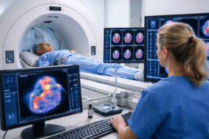
Single-photon emission computed tomography (SPECT) is an advanced nuclear cardiology imaging test used to diagnose and evaluate coronary artery disease and cardiac ischemia (reduced blood flow to the heart muscle). SPECT imaging is commonly performed as part of a nuclear stress test and helps cardiologists assess how well blood is circulating through the heart during rest and stress conditions.
Similar to myocardial perfusion imaging (MPI), the SPECT test begins with the injection of a small amount of a safe radioactive tracer into the bloodstream. This tracer emits gamma rays that are detected by specialized imaging equipment, including a gamma camera and, in many cases, an integrated CT scanner. These technologies work together to produce detailed, three-dimensional images that allow cardiologists to evaluate blood flow patterns and heart muscle function.
SPECT imaging helps identify areas of the heart receiving adequate blood supply, as well as areas affected by blocked or narrowed coronary arteries, scar tissue from prior heart attacks, or regions at risk for future cardiac events.
Cardiac positron emission tomography combined with computed tomography (PET-CT) is another highly advanced nuclear cardiology imaging technique that provides additional diagnostic information beyond traditional SPECT imaging. PET-CT allows cardiologists to obtain highly detailed images of coronary artery structure, measure coronary calcium buildup, and evaluate blood flow to the heart muscle with exceptional accuracy.
PET-CT imaging offers several advantages, including improved image clarity, enhanced detection of coronary artery disease, and precise measurement of myocardial blood flow. These capabilities help cardiologists determine whether patients may benefit from additional procedures such as coronary angiography, stent placement, or other cardiovascular interventions.
Both SPECT and PET-CT imaging are highly effective tools for diagnosing coronary artery disease, evaluating blood supply to damaged areas of the heart, and guiding personalized treatment plans designed to improve long-term cardiovascular outcomes.
What do nuclear cardiology procedures have in common? These advanced diagnostic tests are minimally invasive, highly accurate, and essential tools for the early detection and prevention of heart disease. Nuclear cardiology imaging techniques, including nuclear stress testing, SPECT imaging, and PET-CT cardiac scans, help cardiologists evaluate blood flow to the heart muscle, assess heart function, and identify areas of previous or ongoing heart damage.
Determining how well your heart performs under stress through nuclear stress testing is an important step in diagnosing coronary artery disease and developing an effective, personalized treatment plan. These advanced imaging tests allow cardiologists to detect cardiovascular problems early, often before symptoms become severe, improving long-term heart health outcomes. That’s why, it’s time to consult with a board-certified cardiologist in Tampa, FL, equipped with years of experience in nuclear cardiology.
Patients seeking advanced nuclear cardiology testing and preventative heart care in Tampa, Florida, can trust the experience and expertise of Sachin Diwadkar, MD, at Ascent Cardiology. Dr. Diwadkar is board certified in nuclear cardiology and has extensive experience using advanced cardiac imaging techniques to diagnose and manage coronary artery disease and other cardiovascular conditions.
To consult a cardiologist in Tampa, FL, from Ascent Cardiology, please request an appointment today.
Medical Disclaimer
The content provided on the Ascent Cardiology website is intended for general educational and informational purposes only. The information, articles, and media presented on this website do not constitute medical advice, diagnosis, or treatment recommendations. Content published on this website is not intended to replace consultation, evaluation, or treatment by qualified healthcare professionals, including the providers at Ascent Cardiology or other licensed medical professionals.
If you believe you may be experiencing a medical emergency, call 911 or seek immediate emergency medical care.
The medical information, opinions, and educational materials presented on this website are provided for general awareness and may not reflect the specific clinical practices or medical opinions of Ascent Cardiology or its providers. Reliance on any information provided on this website is solely at your own risk.
This website may include links to third-party medical or educational websites for informational purposes only. Ascent Cardiology does not control, endorse, or assume responsibility for the accuracy, reliability, or content of any third-party websites linked from this site.
How High Blood Pressure Affects Your Heart, Brain, and Body
High blood pressure—also known as hypertension—is one of the most common and dangerous cardiovascular conditions worldwide. Often called the “silent

Stress and Heart Disease: How to Protect and Improve Your Heart Health
Chronic stress is increasingly recognized as a major risk factor for cardiovascular disease. Many high-performing, Type A individuals juggle demanding
What Is Nuclear Cardiology? Tests, Benefits, and What Patients Should Know
Nuclear cardiology is a specialized field of cardiovascular medicine that uses safe, low-dose radioactive tracers combined with advanced imaging technology